


Solis Mammography Discusses Advances in Women’s Health Technology
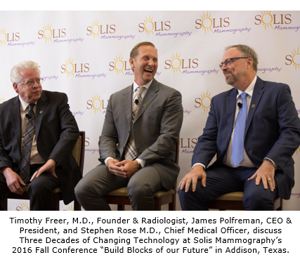
Katy, TX News (February 6, 2017) – In honor of Solis Mammography’s 30th anniversary in 2016, three pioneers in women’s breast health – Dr. Timothy Freer, founder and practicing radiologist; Dr. Stephen Rose, chief medical officer; and James Polfreman, CEO and president of Solis Mammography – gathered for a roundtable discussion.
Describing the origins of what is now Solis Mammography, Dr. Freer remarked, “It’s pretty amazing that what started off as one 600-square-foot location, one radiologist, one film mammography machine with one technologist in Plano, Texas, in 1986 has today grown into Solis Mammography, the nation’s largest independent provider of breast imaging services with 38 centers across six major markets serving more than 600,000 patients each year.”
Although the panel discussion revolved around three decades of serving breast health, more time was spent talking about the changing role of technology in advancing breast imaging – specifically with advances in three-dimensional (3-D) mammography, also known as digital breast tomosynthesis.
Dr. Rose explained that his first exposure to mammography was Xerox, “Blue and white paper was how we interpreted mammograms at that time. The technology of film screening followed, bringing mammogram resolution to an entirely different level than ever seen before. Digital mammography slightly improved film screening but wasn’t very exciting. However, by 2015, over 95 percent of all mammograms were digital.”1
“Film to digital mammography was an evolution. Digital mammography to 3-D mammography is a revolution,” Dr. Rose stated.
From a patient point of view, there is no noticeable difference between 2-D exam and 3-D exam during the mammogram as both take about the same amount of time, compression and positioning. However, from a radiologist point of view, the difference in the images between 2-D and 3-D mammography is night and day. Providing around 60 images of breast tissue, divided into 1 mm slices, 3-D mammography allows the radiologist to see cancers as early as stage zero and to find masses that might otherwise be hidden within dense breast tissue. This compares to conventional 2-D mammography which offers the radiologist just two images of each breast through compressed breast tissue.
To better understand the difference between conventional 2-D and 3-D mammography, imagine a book with clear covers and pages. With 2-D, the radiologist must peer through the front or back cover, looking for a single word that does not have the same characteristics as the other words. With 3-D, the breast is seen in 1mm slices, giving the radiologist the ability to look for that abnormal word one “page” at a time.
Dr. Rose reported that Solis Mammography’s greatest accomplishment in 30 years is “the implementation of 3-D mammography.” Peer-reviewed research, co-authored by Dr. Rose, has shown that 3-D mammography increases early detection of breast cancer by 54 percent and decreases recall rates by 37 percent. (A recall is the radiologist calling the patient back for more tests to ensure accuracy).
“To have our chief medical officer be a principal expert on 3-D technology is uniquely distinctive and a reflection of Solis Mammography’s dedication to offering the highest quality care with exceptionally accurate results,” Polfreman said. “From Dr. Rose’s early research on 3-D, to diagnosing the first patient with bilateral breast cancer that was originally missed using the standard 2-D, to his most recent study that proves 3-D is extremely beneficial to women in their 40s — these are critical advances in the clinical science of mammography allowing Solis to be a leading authority on breast health.”
Solis Mammography’s commitment to 3-D technology has translated to $18 million invested over the past three years to upgrade all of its centers to provide 3-D mammography. To date, 37 of the 38 Solis centers offer 3-D mammography, with the final center being upgraded this month in Chandler, Arizona.
Implementing the best mammography technology is step one, but helping patients afford that technology must follow in suit. Medicare announced full coverage for 3-D mammography in January 2015. Other private insurers have added coverage but only in select regional markets. And CIGNA was the first national private insurer to offer 3-D coverage in all U.S. markets. Solis Mammography is working to support both federal and state legislation which supports a woman’s right to 100% coverage for 3-D mammography. “We want to ensure that a woman’s decision on what type of mammogram she has won’t be determined by her financial means,” commented Polfreman.
Simply put, 3-D mammography is the best and most advanced technology for early detection of breast cancer. Early detection not only saves lives, it improves quality of life – providing women better and less invasive options for treatment. Dr. Rose concluded, “I know without a shadow of a doubt that every patient we see is benefiting from the service we provide.”
To watch Solis Mammography’s 30th anniversary video, Three Decades of Breast Health, go to http://newsroom.solismammo.com/2017/01/09/solis-mammography-30th-anniversary/. To view Solis Mammography’s 3-D Mammography infographic go to: http://newsroom.solismammo.com/2016/06/01/3dinfo/.